

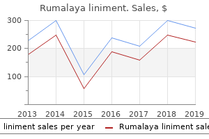
"Purchase rumalaya liniment discount, muscle relaxer 75".
By: X. Goran, M.A., M.D.
Medical Instructor, Washington University School of Medicine
All events flowing right from those points are blocked in cells with mutations in the indicated genes spasms left side under rib cage proven rumalaya liniment 60 ml. The events and gene products shown here are discussed in greater detail in later chapters spasms upper left quadrant cheap rumalaya liniment 60ml with visa. Fission yeast gene names are typically written in lower-case italics spasms or twitches rumalaya liniment 60 ml visa, with a plus sign to signify the wild-type gene (for example cdc2+ for the wild type spasms left shoulder blade cheap rumalaya liniment online visa, cdc2-1 for a mutant allele of that gene). The convention for protein names is less established, but they are generally not italicized and are written with only the first letter capitalized (for example Cdc28); the letter p is sometimes added to signify protein (for example Cdc28p). These simple early divisions also lack many of the checkpoint controls found in somatic cell cycles. As a result, the study of embryonic cell division, particularly in the frog Xenopus laevis, has led to many important insights into the fundamental logic and components of cell-cycle control. Fertilization of the Xenopus egg triggers a remarkably rapid and synchronous series of 12 cleavage divisions (Figure 2-9; see also Figure 2-2). These rapid divisions lack gap phases and quickly subdivide the egg into the blastula, a ball of 4,000 cells. Gap phases appear in the cell cycle, cell growth occurs and the cell-cycle control system begins to assume its more complex adult form. Studies of cell division in the pre-blastula Xenopus embryo were the first to suggest the existence of an autonomous cell-cycle oscillator, or clock, that continues to operate with normal timing even if cell-cycle events are severely crippled by removal of the nucleus (Figure 2-10). This oscillator is not readily apparent in most other cell types, in which checkpoint mechanisms arrest the clock when cell-cycle events fail to occur. Figure 2-9 Early embryogenesis in Xenopus Fertilization of the mature egg initiates rapid cleavage divisions, which subdivide the cytoplasm into smaller and smaller cells until the embryo reaches the blastula stage (see Figure 2-2). The complex morphogenetic movements of gastrulation, or formation of the gut, then occur, and the embryo is quickly transformed into a free-swimming tadpole. Unfertilized eggs develop from diploid oocytes by meiosis the Xenopus egg is derived from a much smaller cell known as an oocyte. Soon after its birth in the ovary, the diploid oocyte enters the meiotic program and completes meiotic S phase (see Chapter 9 for a description of the stages of meiosis). It then arrests in meiotic prophase for several months, during which it grows to a diameter of about 1 mm. In response to hormonal cues from the pituitary gland, the follicle cells surrounding the oocyte then secrete the hormone progesterone, which interacts with the oocyte to initiate oocyte maturation. At division, one chromosome set remains in the oocyte and the other is discarded in the polar body, which is eventually resorbed. The oocyte has now been converted to an unfertilized egg and maturation is complete. This is followed by the fusion of the haploid egg nucleus with the haploid sperm nucleus, and the resulting diploid zygote begins the first mitotic cell division of the early embryo. Because of their large size, it is possible to inject Xenopus oocytes, eggs and early embryos with various test substances. The early embryonic cell cycle can be reconstituted in a test tube height, millimeters 1 0. Mock fertilization, or activation, of these eggs can be achieved with electrical stimulation, which triggers a sudden influx of calcium ions into the eggs. Gentle centrifugation of activated frog eggs breaks them apart and stratifies their contents, allowing the isolation of essentially undiluted egg cytoplasm. When Xenopus sperm nuclei stripped of their membranes are added to this cytoplasm in a test tube, the sperm chromosomes decondense and are packaged in a nuclear envelope. This cycle of S and M phases can repeat itself in the test tube for several rounds, with cell-cycle lengths about twice that of a normal embryonic cell cycle. The ability to reconstruct an early embryonic cell cycle in a test tube provides an unparalleled system for the biochemical dissection of basic cell-cycle control. Foreign proteins, chemicals or inhibitory antibodies can be added to these extracts to test their effects on cell-cycle progression. In the non-nucleated half, divisions do not occur, but the surface of the egg exhibits periodic contractions, resulting in the indicated changes in the height of the non-nucleated fragment.
We also wish to thank the Open Learning Initiative at Carnegie Mellon University back spasms 7 weeks pregnant proven rumalaya liniment 60ml, with whom we shared and exchanged resources during the development of Human Anatomy and Physiology spasms in your sleep cheap rumalaya liniment 60 ml without a prescription. At some point in the future spasms video cheap rumalaya liniment uk, will this type of technology lead to the ability to augment our nervous systems That quote is from the early 1990s; in the two decades since muscle relaxant liver disease cheap 60 ml rumalaya liniment with mastercard, progress has continued at an amazing rate within the scientific disciplines of neuroscience. It is an interesting conundrum to consider that the complexity of the nervous system may be too complex for it (that is, for us) to completely unravel. One easy way to begin to understand the structure of the nervous system is to start with the large divisions and work through to a more in-depth understanding. In other chapters, the finer details of the nervous system will be explained, but first looking at an overview of the system will allow you to begin to understand how its parts work together. The focus of this chapter is on nervous (neural) tissue, both its structure and its function. But before you learn about that, you will see a big picture of the system-actually, a few big pictures. That suggests it is made of two organs-and you may not even think of the spinal cord as an organ-but the nervous system is a very complex structure. Within the brain, many different and separate regions are responsible for many different and separate functions. It is as if the nervous system is composed of many organs that all look similar and can only be differentiated using tools such as the microscope or electrophysiology. In comparison, it is easy to see that the stomach is different than the esophagus or the liver, so you can imagine the digestive system as a collection of specific organs. The Central and Peripheral Nervous Systems the nervous system can be divided into two major regions: the central and peripheral nervous systems. The brain is contained within the cranial cavity of the skull, and the spinal cord is contained within the vertebral cavity of the vertebral column. In actuality, there are some elements of the peripheral nervous system that are within the cranial or vertebral cavities. The peripheral nervous system is so named because it is on the periphery-meaning beyond the brain and spinal cord. Depending on different aspects of the nervous system, the dividing line between central and peripheral is not necessarily universal. A glial cell is one of a variety of cells that provide a framework of tissue that supports the neurons and their activities. The neuron is the more functionally important of the two, in terms of the communicative function of the nervous system. To describe the functional divisions of the nervous system, it is important to understand the structure of a neuron. Neurons are cells and therefore have a soma, or cell body, but they also have extensions of the cell; each extension is generally referred to as a process. There is one important process that every neuron has called an axon, which is the fiber that connects a neuron with its target. Looking at nervous tissue, there are regions that predominantly contain cell bodies and regions that are largely composed of just axons. These two regions within nervous system structures are often referred to as gray matter (the regions with many cell bodies and dendrites) or white matter (the regions with many axons). The colors ascribed to these regions are what would be seen in "fresh," or unstained, nervous tissue. It can be pinkish because of blood content, or even slightly tan, depending on how long the tissue has been preserved. But white matter is white because axons are insulated by a lipid-rich substance called myelin. Lipids can appear as white ("fatty") material, much like the fat on a raw piece of chicken or beef. Actually, gray matter may have that color ascribed to it because next to the white matter, it is just darker-hence, gray.
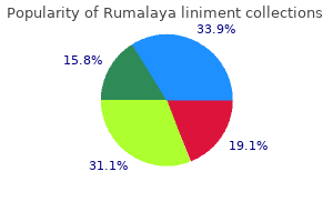
Best buy for rumalaya liniment. Cupping Therapy for Muscle Relaxation Neck and Shoulder Pain Santa Monica Acupuncture.
The diencephalon is between the cerebrum and the rest of the nervous system and can be described as the region through which all projections have to pass between the cerebrum and everything else spasms back pain and sitting purchase rumalaya liniment 60 ml visa. The brain stem includes the midbrain spasms thumb joint rumalaya liniment 60 ml online, pons muscle relaxant 10mg order 60ml rumalaya liniment otc, and medulla muscle relaxant homeopathy buy rumalaya liniment 60ml without a prescription, which correspond to the mesencephalon, metencephalon, and myelencephalon. The cerebellum, being a large portion of the brain, is considered a separate region. One other benefit of considering embryonic development is that certain connections are more obvious because of how these adult structures are related. The retina, which began as part of the diencephalon, is primarily connected to the diencephalon. The eyes are just inferior to the anterior-most part of the cerebrum, but the optic nerve extends back to the thalamus as the optic tract, with branches into a region of the hypothalamus. There is also a connection of the optic tract to the midbrain, but the mesencephalon is adjacent to the diencephalon, so that is not difficult to imagine. The cerebellum originates out of the metencephalon, and its largest white matter connection is to the pons, also from the metencephalon. There are connections between the cerebellum and both the medulla and midbrain, which are adjacent structures in the secondary vesicle stage of development. In the adult brain, the cerebellum seems close to the cerebrum, but there is no direct connection between them. The four ventricles and the tubular spaces associated with them can be linked back to the hollow center of the embryonic brain (see Table 13. Stages of Embryonic Development Neural tube Anterior neural tube Anterior neural tube Table 13. A groove forms along the dorsal surface of the embryo, which becomes deeper until its edges meet and close off to form the tube. If this fails to happen, especially in the posterior region where the spinal cord forms, a developmental defect called spina bifida occurs. The closing of the neural tube is important for more than just the proper formation of the nervous system. There are three classes of this disorder: occulta, meningocele, and myelomeningocele (Figure 13. The first type, spina bifida occulta, is the mildest because the vertebral bones do not fully surround the spinal cord, but the spinal cord itself is not affected. No functional differences may be noticed, which is what the word occulta means; it is hidden spina bifida. The other two types both involve the formation of a cyst-a fluid-filled sac of the connective tissues that cover the spinal cord called the meninges. The earlier that surgery can be performed, the better the chances of controlling or limiting further damage or infection at the opening. For many children with meningocele, surgery will alleviate the pain, although they may experience some functional loss. Because the myelomeningocele form of spina bifida involves more extensive damage to the nervous tissue, neurological damage may persist, but symptoms can often be handled. Complications of the spinal cord may present later in life, but overall life expectancy is not reduced. The result is the emergence of meninges and neural tissue through the vertebral column. The spinal cord is a single structure, whereas the adult brain is described in terms of four major regions: the cerebrum, the diencephalon, the brain stem, and the cerebellum. The coordination of reflexes depends on the integration of sensory and motor pathways in the spinal cord. The Cerebrum the iconic gray mantle of the human brain, which appears to make up most of the mass of the brain, is the cerebrum (Figure 13. The wrinkled portion is the cerebral cortex, and the rest of the structure is beneath that outer covering.
The parasite then rapidly penetrates into the cytosol and differentiates into the amastigote stage muscle relaxant you mean whiskey purchase generic rumalaya liniment line. After several division cycles spasms between ribs purchase rumalaya liniment with american express, some of the parasites transform back into trypomastigotes spasms from dehydration buy generic rumalaya liniment on line. The affected cells die spasms of the larynx order genuine rumalaya liniment, releasing the parasites that can now enter the bloodstream and become distributed throughout the body. They infect cells in many types of tissues, including the central nervous system, heart muscle, the myenteric plexus, the urogenital tract, and the reticuloendothelial system. Thousands of organisms are produced within one insect without apparently affecting it. Epimastigotes maintain their place in the gut of the insect by specific receptorligand interactions involving at least one parasite surface glycoprotein and a carbohydrate lectin on the gut cells of the insect. Cellular and Molecular Pathogenesis Infection with Trypanosoma cruzi results in partial immunosuppression that further aids the parasite in remaining inside the host cell for extended periods of time. The surface coat of the free-swimming trypomastigote contains a specific complement regulatory protein that binds the C3b and C4b components, inhibiting the alternate pathway. Infected individuals remain infected for life and most of the pathological consequences result from cell death. Myenteric plexus damage results in loss of muscle tone and enlargement of the organ, particularly the digestive tract. It is thought that most of the damage to the myenteric plexus is directly resultant on damage that occurs early in infection and is not thought to be prevented by treatment of chronic Chagas. Current thinking regarding a dominant role for auto-antibodies inducing cardiomyopathy plays down this mechanism to account for heart damage during chronic infection. In acute Chagas disease acquired from the kissing bug, the incubation period is from a few days to two weeks before the onset of symptoms. Severe acute manifestations are estimated to develop in approximately 1% of cases and may involve acute myocarditis, pericardial effusion, and in some cases meningoencephalitis. The swollen eyelid is firm to the touch, and there may be associated conjunctivitis. If the bite occurs elsewhere, the adjacent area is erythematous, brawny, and firm to the touch. When the chagoma disappears after several weeks, it leaves an area of depigmentation. An associated neuropathy develops, and then disappears when the patient enters the chronic phase of the infection. Chronic Chagas Disease Most patients survive the acute phase and become asymptomatic. Patients at this point enter the chronic phase and although they may remain symptomatic they are capable of transmitting parasites to the insect vector. During the chronic phase of Chagas disease individuals enter an indeterminant or latent stage, which may continue for the life of most (70-80%) affected individuals, or which may later manifest with injury to the heart or gastrointestinal system. In as many as a third of patients, there is a progression of the disease that usually manifests as cardiac or gastrointestinal disease. Ultrastructural studies showed that vinculin costameres in cardiomyocytes become disrupted during intracellular infection with the amastiogote stage, and this is thought to make a major contribution to the cardiomyopathy so typically seen in the chronic infection. Gastrointestinal involvement mainly manifests with the development of megaesophagus, characterized by dysphagia and regurgitation, and megacolon, leading to constipation and fecal retention. Rarely it also leads to megaureters, megabladder, megagallbladder, and bronchiectasis. It is estimated that 1-10% of infants born to an infected mother will acquire congenital Chagas disease. In acute disease the level of parasites in the blood is high enough that trypomastigotes can be detected on examination of Giemsa-stained blood smears, and both a thin and thick smear should be ordered. This screening test is not a diagnostic test and would not count as one of the two diagnostic test results. Inoculating blood from suspected individuals into susceptible animals can reveal the organism, but this approach presents too much impracticality for most diagnostic facilities. Xenodiagnosis, employing uninfected kissing bugs, allowing them to feed on the patient, then dissecting the bugs some days later, can also reveal the presence of parasites in chronically infected individuals, but it is a special test requiring extensive laboratory infrastructure and technical assistance. Also prolonged treatments are required lasting 1-2 months, and many patients (as many as one in five) cannot tolerate full treatment courses. Additional new and sobering information indicates that once heart involvement begins, it may be too late for the drugs to have a clinical impact.
